This article aims to report a rare case of a 38 years old Para 2 who was referred to the outpatient clinic of Vikash Multispecialty Hospital as a case of grade 3 uterovaginal prolapse with complaint of something coming out per vagina since 12 years. She also complained of heavy bleeding during menstruation, not responding to medical management since 2 years. Clinical and imaging studies lead to the diagnosis of anterior vaginal cyst, intramural fibroid and cervical polyp. After detailed counseling of the risks and benefits of surgical excision of the cyst with or without removal of uterus, she opted for removal of vaginal cyst and vaginal hysterectomy as she was tired of the medical management and had to undergo blood transfusion twice due to heavy menstrual bleeding. She had an uneventful postoperative period and was discharged in good condition on the third day. This case stresses on the importance of right diagnosis and proper management of the masses coming out of vagina as not everything which comes out of vagina is prolapse. Hesitancy in seeking medical care and lack of proper medical facilities, especially in rural areas in underdeveloped and developing countries, are among the major reasons these cases are unreported in medical literature.
This is an Open Access article, distributed under the terms of the Creative Commons Attribution 4.0 International License (http://creativecommons.org/licenses/by/4.0/), which permits unrestricted use, distribution and reproduction in any medium or format, provided the original work is properly cited.
Vaginal cysts are rare, benign, predominantly cystic lesions, commonly of the anterior vaginal wall. They typically present in 3rd or 4th decade of life and also seen in children and postmenopausal women
[1]
R. N. Marisa and E. Oliva, Gynecologic Pathology, Elsevier Health Sciences, Philadelphia, Pa, USA, 2009.
[2]
Junaid TA, Thomas SM. Cysts of the vagina and vulva: a comparative study. Int J GynecolObstet 1981; 19: 239-243.
[1, 2]
. Vaginal cysts are usually small (0.5-2 cm in diameter), solitary and asymptomatic; however they can increase in size and are often misdiagnosed as anterior vaginal prolapse. Therefore, complete surgical vaginal excision of the symptomatic vaginal lesion is feasible and constitutes a good management option
[3]
Tsiapakidou S, Theodoulidis I, Grimbizis G, Mikos T. Surgical excision of vaginal cysts presenting as pelvic organ prolapse: a case series. Pan Afr Med J. 2022 May 6; 42: 10.
Mrs A, 38 years old, Para 2 was referred to the outpatient clinic of Vikash Multispecialty Hospital as a case of grade 3 uterovaginal prolapse with complaint of something coming out per vagina since 12 years and heavy bleeding during menstruation, not responding to medical management since 2 years. She had developed anemia due to heavy menstrual bleeding and had to undergo blood transfuison twice.
On examination, patient was pale. Abdomen was soft with no palpable masses.
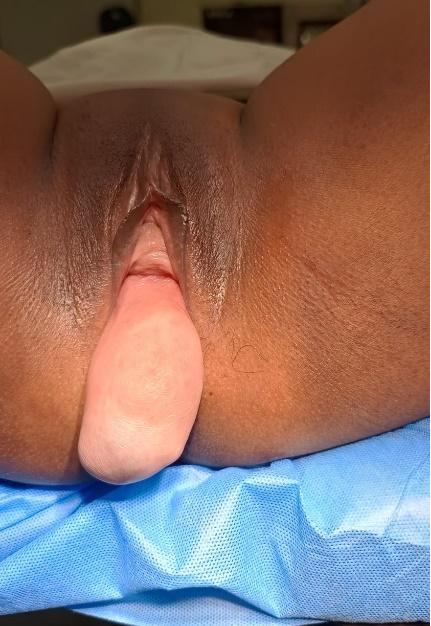
Perineal inspection appeared as grade 3 anterior vaginal wall prolapse but close examination after urethral catherization showed minimal cystocoele with large anterior vaginal cyst (Figure 1). The cyst was about 8 x 7 cm in size. It appeared extending from the upper part of right lateral fornix with the lower edge at the cervicovaginal junction.
Per speculum examination showed a fleshy structure protruding through the cervical os.
Uterus was enlarged to 8 weeks size on bimanual examination with a firm growth protruding through the os. Growth was felt free from the endocervical canal and upper edge was not reached.
Ultrasound showed enlarged uterus with a 2.5 x 1.4 cm sized well defined hypoechoic mass lesion seen in the anterior wall of uterus and another well defined hyper echoic lesion measuring 3 x 2.4 cm in the cervical region with well defined vascular pedicle but no details was mentioned about the vaginal cyst.
Contrast CT scan was done which showed a fluid filled globular structure of size 7.9 x 4.8 x 3.3 cm projecting outside the vulva from the vaginal canal. There was no evidence of filling of contrast in the delayed phases within the cystic lesion protruding from the vulva. Cervix and uterus were stretched inferiorly till just above the introitus.
The patient was counseled about the findings.
She wanted definitive management of her clinical condition of heavy menstrual bleeding and vaginal cyst.
After detailed counseling of the risks and benefits, she opted for removal of vaginal cyst and vaginal hysterectomy.
She received 1 unit of blood preoperatively in view of anemia.
Small incision was given in the lower part of the vaginal cyst and extended superiorly. The cyst got ruptured during enucleation and mucinous brownish fluid was drained. Cyst wall was dissected and redundant vaginal walls were excised. Cavity was closed with Vicryl no. 1. Removal of uterus and cervix was done by vaginal route. Bilateral tubes and ovaries were preserved. Vaginal vault was closed with Vicryl no 1 after haemostasis.
Cut Specimen was noted to have 4 x 4 cm intramural fibroid with sub mucosal component which was protruding through the os.
Histopathogical examination of the specimen showed vaginal cyst lined by ciliated columnar epithelium and was labeled as Mullerian cyst. Full report of the specimen included Chronic papillary cervicitis, Endometrial polyp, Leiomyoma no signs of atypia or malignancy.
Her postoperative stay was uneventful and she was discharged home on the third postoperative day.
3. Discussion
Vaginal cysts have an estimated prevalence of 0.5-1%. Being rare, these pose as a diagnostic challenge upon presentation
[1]
R. N. Marisa and E. Oliva, Gynecologic Pathology, Elsevier Health Sciences, Philadelphia, Pa, USA, 2009.
[1].
Their origin may be Mullerian (paramesonephric), Wolffian (mesonephric), squamous (traumatic), or urogenital. The cysts are usually small, ranging from 0.1 to 2 cm in diameter, although they may measure >4 cm. The differential diagnoses of a cyst in the lower female genital tract include Mullerian cyst, inclusion ¨ cyst, mesonephric cyst (Gartner’s duct), Bartholin gland cyst, urethrocele, urethral diverticulum, Skene’s duct cyst, pelvic organ prolapse, hematocolpos, and a myxomatous tumour
[4]
S. G. Fletcher and G. E. Lemack, “Benign masses of the female periurethral tissues and anterior vaginal wall,” Current Urology Reports, vol. 9, no. 5, pp. 389–396, 2008.
[4]
. Simple mesonephric (Gartner’s) or paramesonephric (Mullerian) cysts may occur at especially high levels near the fornices.
40% of the cystic vaginal masses of embryologic origin are Müllerian cysts
[7]
Hwang JH, Oh MJ, Lee NW, Hur JY, Lee KW, et al. (2009) Multiple vaginal mullerian cysts: a case report and review of literature. Arch Gynecol Onstet 280(1): 137-139.
[8]
Eilber KS, Raz S (2003) Begnign cystic lesions of the vagina: a literature review. J Urol 170(3): 717-722.
[7, 8]
Gartner’s duct cysts are less common than Mullerian ¨ cysts, comprising approximately 10% of benign vaginal cysts.
The prevalence might be under reported due to hesitancy in seeking medical care especially in rural areas in developing countries. The above mentionedcase finally presented at 38 years of age with her chief complaint of heavy menstrual bleeding though she was suffering for the past 12 years with dyspareunia, difficulty in walkingdue to the vaginal cyst.
Müllerian cysts arise from persistence of pseudostratified columnar epithelium during its replacement with squamous epithelium. This epithelium can persist anywhere in the vagina but Müllerian cysts are generally located anterolaterally near the vaginal fornix
[5]
Lallar M, Nandal R, Sharma D, Shastri S (2015) Large posterior vaginal cyst in pregnancy. BMJ Case Report 2015.
[6]
Arumugam AV, Kumar G, Si LK, Vijayananthan A (2007) Gartner duct cyst in pregnancy presenting as a prolapsing pelvic mass. Biomed Imaging Interv J 3(4): e46.
[8]
Eilber KS, Raz S (2003) Begnign cystic lesions of the vagina: a literature review. J Urol 170(3): 717-722.
[5, 6, 8]
. Their size varies generally between 1cm to 7cm but it can sometimes be much bigger. 40% of the cystic vaginal masses of embryologic origin are Müllerian cysts
[8]
Eilber KS, Raz S (2003) Begnign cystic lesions of the vagina: a literature review. J Urol 170(3): 717-722.
[10]
Prdhan S, Tobon H (1986) Vaginal cysts: a clinicalpathological study of 41 cases. Int J Gynecol Pathol 5(1): 35-46.
[8, 10]
. Dyspareunia, pelvic pain, pressure, bulging mass and urinary incontinence or obstruction are the most frequent presenting symptoms associated with vaginal cyst
[8]
Eilber KS, Raz S (2003) Begnign cystic lesions of the vagina: a literature review. J Urol 170(3): 717-722.
[8]
.
Mullerian and Gartner cysts are benign tumors, but malignancy must be considered. Lee reported adenocarcinoma arising in a Mullerian vaginal cyst
[9]
Lee KS, Park KH, Lee S, Kim JY, Seo SS (2005) Adenocarcinoma arising in a vaginal mullerian cyst: a case report. Gynecol Oncol 99(3): 767-769.
[9]
. In literature there are very rare reported cases of malignant vaginal cysts, but even with an asymptomatic and small cyst this eventuality should be considered.
Cysts should be characterized by radiologic investigations, Ultrasound and computed tomography (CT) are helpful to define the cystic mass
[7]
Hwang JH, Oh MJ, Lee NW, Hur JY, Lee KW, et al. (2009) Multiple vaginal mullerian cysts: a case report and review of literature. Arch Gynecol Onstet 280(1): 137-139.
[11]
Tiwari U, Relia N, Shailesh F, Kaushik C (2014) Gartner Duct Cyst: CT and MRI findings. J Obstet Gynaecol India 64(Suppl 1): 150-151.
[12]
Elsayes KM, Narra VR, Dillman JR, Velcheti V, Hameed O, et al. (2007) Vaginal masses: magnetic Resonance Imaging features with Pathologic correlation. Acta Radiol 48(8): 921-933.
[7, 11, 12]
. Magnetic Resonance Imaging (MRI) is the gold standard to characterize surrounding tissues and define origin and extent of the pelvic mass. These cysts have been observed in 1 to 2% of female pelvic MRI examinations
In our case, we did contrast CT due to low cost keeping in mind the socio-economic condition of the patient. The radiologist described a globular cystic lesion prolapsing through the vulva stretching the cervix till above the introitus with no evidence of filling of the contrast in the delayed phase within the cystic lesion. Use of MRI might have allowed better characterization of the müllerian vaginal cyst as MRI affords excellent visualisation of the vagina and surrounding tissue, offering high-contrast resolution and multiplanar capabilities
[16]
Santos X. M., Krishnamurthy R., Bercaw-Pratt J. L., and Dietrich J. E., The utility of ultrasound and magnetic resonance imaging versus surgery for the characterization of Müllerian anomalies in the pediatric and adolescent population, Journal of Pediatric & Adolescent Gynecology. (2012) 25, no. 3, 181–184,
. Retrograde urography into the cystic lesion permits the investigation of the proximity of the urinary tract to the cyst.
Full surgical excision constitutes a good management option. As the margins of the cyst were well defined on vaginal examination, we took the vaginal route to excise the cyst which also facilitated the vaginal hysterectomy by creating space for manipulation of the uterus.
Vaginal cysts usually do not necessitate medical intervention as they may not cause symptoms. Salete et al had followed up 4 patients of age group (37-53 years) for 2 to 17 years.; no indication for operative management appeared during that time
[15]
Rios, S. S., Pereira, L. C. R., Santos, C. B. et al. Conservative treatment and follow-up of vaginal Gartner’s duct cysts: a case series. J Med Case Reports 10, 147 (2016).
[15]
. The management of the symptomatic vaginal cysts is primarily surgical.
[14]
Boujenah J, Ssi-Yan-Kan G, Prevot S, Chalouhi GE, Deffieux X. A vaginal Gartner duct cyst presenting as a cystocele during pregnancy. Eur J ObstetGynecolReprod Biol. 2014 Sep; 180: 202–4.
[14]
. Many authors have proposed marsupialization of the cyst, and it seems that it is a safe, minimally invasive technique with satisfactory long-term results
[14]
Boujenah J, Ssi-Yan-Kan G, Prevot S, Chalouhi GE, Deffieux X. A vaginal Gartner duct cyst presenting as a cystocele during pregnancy. Eur J ObstetGynecolReprod Biol. 2014 Sep; 180: 202–4.
[14]
. The size of the cyst may cause difficulties during its excision, and a combined approach (abdominal-perineal or laparoscopic-perineal) may be adopted by the surgeons in order to secure its complete removal.
4. Conclusion
Cystic lesions of the vagina are a common occurrence in women in their third and fourth decades, and represent a spectrum of disease from embryological derivatives to preneoplastic lesions
[17]
KARYN SCHLUNT EILBER, SHLOMO RAZ, Benign Cystic Lesions of the Vagina: A Literature Review, The Journal of Urology, Volume 170, Issue 3, 2003, Pages 717-722, ISSN 0022-5347,
. Mainly these are small in size and asymptomatic. Large symptomatic vaginal cysts can present as genital prolapse and may be difficult to diagnose. When evaluating an anterior vaginal cyst, assessment of the lesion via history taking and pelvic examination is important to confirm lesion size and location. Imaging by ultrasound or MRI is essential for diagnosis.
[16]
Santos X. M., Krishnamurthy R., Bercaw-Pratt J. L., and Dietrich J. E., The utility of ultrasound and magnetic resonance imaging versus surgery for the characterization of Müllerian anomalies in the pediatric and adolescent population, Journal of Pediatric & Adolescent Gynecology. (2012) 25, no. 3, 181–184,
The surgical excision of symptomatic lesion is the preferred modality of treatment with a favorable outcome for the patients.
Abbreviations
MRI
Magnetic Resonance Imaging
CT
Computed Tomography
Author Contributions
Sweety Kumari is the sole author. The author read and approved the final manuscript.
Conflicts of Interest
The author declares no conflicts of interest.
References
[1]
R. N. Marisa and E. Oliva, Gynecologic Pathology, Elsevier Health Sciences, Philadelphia, Pa, USA, 2009.
[2]
Junaid TA, Thomas SM. Cysts of the vagina and vulva: a comparative study. Int J GynecolObstet 1981; 19: 239-243.
[3]
Tsiapakidou S, Theodoulidis I, Grimbizis G, Mikos T. Surgical excision of vaginal cysts presenting as pelvic organ prolapse: a case series. Pan Afr Med J. 2022 May 6; 42: 10.
S. G. Fletcher and G. E. Lemack, “Benign masses of the female periurethral tissues and anterior vaginal wall,” Current Urology Reports, vol. 9, no. 5, pp. 389–396, 2008.
[5]
Lallar M, Nandal R, Sharma D, Shastri S (2015) Large posterior vaginal cyst in pregnancy. BMJ Case Report 2015.
[6]
Arumugam AV, Kumar G, Si LK, Vijayananthan A (2007) Gartner duct cyst in pregnancy presenting as a prolapsing pelvic mass. Biomed Imaging Interv J 3(4): e46.
[7]
Hwang JH, Oh MJ, Lee NW, Hur JY, Lee KW, et al. (2009) Multiple vaginal mullerian cysts: a case report and review of literature. Arch Gynecol Onstet 280(1): 137-139.
[8]
Eilber KS, Raz S (2003) Begnign cystic lesions of the vagina: a literature review. J Urol 170(3): 717-722.
[9]
Lee KS, Park KH, Lee S, Kim JY, Seo SS (2005) Adenocarcinoma arising in a vaginal mullerian cyst: a case report. Gynecol Oncol 99(3): 767-769.
[10]
Prdhan S, Tobon H (1986) Vaginal cysts: a clinicalpathological study of 41 cases. Int J Gynecol Pathol 5(1): 35-46.
[11]
Tiwari U, Relia N, Shailesh F, Kaushik C (2014) Gartner Duct Cyst: CT and MRI findings. J Obstet Gynaecol India 64(Suppl 1): 150-151.
[12]
Elsayes KM, Narra VR, Dillman JR, Velcheti V, Hameed O, et al. (2007) Vaginal masses: magnetic Resonance Imaging features with Pathologic correlation. Acta Radiol 48(8): 921-933.
Boujenah J, Ssi-Yan-Kan G, Prevot S, Chalouhi GE, Deffieux X. A vaginal Gartner duct cyst presenting as a cystocele during pregnancy. Eur J ObstetGynecolReprod Biol. 2014 Sep; 180: 202–4.
[15]
Rios, S. S., Pereira, L. C. R., Santos, C. B. et al. Conservative treatment and follow-up of vaginal Gartner’s duct cysts: a case series. J Med Case Reports 10, 147 (2016).
[16]
Santos X. M., Krishnamurthy R., Bercaw-Pratt J. L., and Dietrich J. E., The utility of ultrasound and magnetic resonance imaging versus surgery for the characterization of Müllerian anomalies in the pediatric and adolescent population, Journal of Pediatric & Adolescent Gynecology. (2012) 25, no. 3, 181–184,
KARYN SCHLUNT EILBER, SHLOMO RAZ, Benign Cystic Lesions of the Vagina: A Literature Review, The Journal of Urology, Volume 170, Issue 3, 2003, Pages 717-722, ISSN 0022-5347,
Kumari, S. (2024). Rare Case of Anterior Vaginal Cyst Presenting as Huge Cystocoele. World Journal of Medical Case Reports, 5(2), 23-26. https://doi.org/10.11648/j.wjmcr.20240502.12
Kumari, S. Rare Case of Anterior Vaginal Cyst Presenting as Huge Cystocoele. World J. Med. Case Rep.2024, 5(2), 23-26. doi: 10.11648/j.wjmcr.20240502.12
@article{10.11648/j.wjmcr.20240502.12,
  author = {Sweety Kumari},
  title = {Rare Case of Anterior Vaginal Cyst Presenting as Huge Cystocoele
},
  journal = {World Journal of Medical Case Reports},
  volume = {5},
  number = {2},
  pages = {23-26},
  doi = {10.11648/j.wjmcr.20240502.12},
  url = {https://doi.org/10.11648/j.wjmcr.20240502.12},
  eprint = {https://article.sciencepublishinggroup.com/pdf/10.11648.j.wjmcr.20240502.12},
  abstract = {This article aims to report a rare case of a 38 years old Para 2 who was referred to the outpatient clinic of Vikash Multispecialty Hospital as a case of grade 3 uterovaginal prolapse with complaint of something coming out per vagina since 12 years. She also complained of heavy bleeding during menstruation, not responding to medical management since 2 years. Clinical and imaging studies lead to the diagnosis of anterior vaginal cyst, intramural fibroid and cervical polyp. After detailed counseling of the risks and benefits of surgical excision of the cyst with or without removal of uterus, she opted for removal of vaginal cyst and vaginal hysterectomy as she was tired of the medical management and had to undergo blood transfusion twice due to heavy menstrual bleeding. She had an uneventful postoperative period and was discharged in good condition on the third day. This case stresses on the importance of right diagnosis and proper management of the masses coming out of vagina as not everything which comes out of vagina is prolapse. Hesitancy in seeking medical care and lack of proper medical facilities, especially in rural areas in underdeveloped and developing countries, are among the major reasons these cases are unreported in medical literature.
},
 year = {2024}
}
TY  - JOUR
T1  - Rare Case of Anterior Vaginal Cyst Presenting as Huge Cystocoele

AU  - Sweety Kumari
Y1  - 2024/12/19
PY  - 2024
N1  - https://doi.org/10.11648/j.wjmcr.20240502.12
DO  - 10.11648/j.wjmcr.20240502.12
T2  - World Journal of Medical Case Reports
JF  - World Journal of Medical Case Reports
JO  - World Journal of Medical Case Reports
SP  - 23
EP  - 26
PB  - Science Publishing Group
SN  - 2994-726X
UR  - https://doi.org/10.11648/j.wjmcr.20240502.12
AB  - This article aims to report a rare case of a 38 years old Para 2 who was referred to the outpatient clinic of Vikash Multispecialty Hospital as a case of grade 3 uterovaginal prolapse with complaint of something coming out per vagina since 12 years. She also complained of heavy bleeding during menstruation, not responding to medical management since 2 years. Clinical and imaging studies lead to the diagnosis of anterior vaginal cyst, intramural fibroid and cervical polyp. After detailed counseling of the risks and benefits of surgical excision of the cyst with or without removal of uterus, she opted for removal of vaginal cyst and vaginal hysterectomy as she was tired of the medical management and had to undergo blood transfusion twice due to heavy menstrual bleeding. She had an uneventful postoperative period and was discharged in good condition on the third day. This case stresses on the importance of right diagnosis and proper management of the masses coming out of vagina as not everything which comes out of vagina is prolapse. Hesitancy in seeking medical care and lack of proper medical facilities, especially in rural areas in underdeveloped and developing countries, are among the major reasons these cases are unreported in medical literature.

VL  - 5
IS  - 2
ER  -